


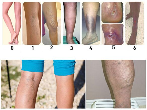
Varicose veins (varicose veins) are a condition in which superficial veins become enlarged or swollen. In most cases, the disease occurs in people over the age of 30. In the vast majority of cases, it is observed in the lower extremities. Varicose veins are characterized by dilation of the lumen of the veins, along with changes in the walls of the veins. The saphenous vein is well contoured and the direction of its course becomes "serpentine". The great saphenous veins are usually involved, the smaller saphenous veins are less frequently involved, and their anastomosis of the great saphenous vein is less common.
causes of varicose veins
Theories proposed to explain the causes and mechanisms of disease onset can be grouped into three groups.
The first group of theories explained the origin of varicose veins through the anatomical features of the location and structure of these vessels in the lower extremities. The veins have valves that prevent the centrifugal flow of blood, which prevents excessive flow of blood from under the skin into the deep veins of the legs. Due to insufficient valves in the saphenous veins, more blood is deposited, causing them to dilate.
A second group of theories on the development of varicose veins value blood stagnation in the pelvis during pregnancy, constipation, the consequences of inflammatory processes, and during prolonged periods in the legs.
The theory of the third group, explaining the origin of varicose veins through constitutional susceptibility, weakness of the mesenchyme, received the least confirmation.
With varicose veins, for various reasons, their walls change and become thinner, so the increased pressure causes the walls to swell. It manifests first in the form of nodes, and at the same time, areas of compaction caused by overgrowth of connective tissue are also noted. Mechanical factors only contribute to the development of venous pathological processes, but are by no means the focus of the pathogenesis, etiology, and causes of lower extremity varicose veins.
Symptoms of varicose veins
As the veins dilate, patients often experience a feeling of fullness and heaviness in the lower extremities. Sometimes there is brief, cramping pain. There is often swelling. Feelings of fullness and heaviness in the extremities increase at night, when edema usually increases. Itching occurs and the legs are often scratched. In the later stages of the disease, ulcers form, usually on the lower third of the inner calf.
The main objective symptom of the disease is visible varicose veins. The patient is examined in a standing position to identify this symptom. At the same time, dilated saphenous veins are clearly visible; on the calf, they appear more prominent and more complex; on the thigh, the veins usually dilate only along the line of the main vascular trunk. Sometimes varicose veins in the thigh are almost at the junction of the largest saphenous vein and the femoral vein. This nodule may be mistaken for a femoral hernia, but the flexibility of the nodule, rapid congestion after removing the examiner's hand, and the presence of dilated veins in the lower leg provide the basis for a correct diagnosis.
There are a number of symptoms that suggest the presence of venous trunk dilation of the great saphenous vein. These include symptoms in which the patient is placed in a horizontal position with the legs elevated. Drain the subcutaneous venous system by carefully stroking the leg from the periphery to the center, apply firm finger pressure to the point where the largest saphenous vein flows into the femoral vein, and hold the finger, and transfer the patient to a standing position. If the venous filling occurs only after the finger is removed, this is a positive symptom. In this case, the anastomosis between the superficial and deep venous networks is poorly expressed and surgery may have a positive impact. If in the patient's vertical position, the surrounding veins still begin to fill slowly, which indicates a significant development of anastomosis - a negative symptom. In this case, the vein ligation procedure will not be successful.
Delbe-Perthes symptoms indicate the extent to which the saphenous vein empties through the anastomosis to the deep vein. The patient is placed with an elastic bandage on the border of the middle and lower third of the thigh, and they are allowed to ambulate slightly. If the tension of the dilated veins is significantly reduced, this indicates a well-developed anastomosis between the superficial and deep veins.
Other symptoms of varicose veins include swelling, eczematous skin changes, and ulcers. Puffiness is different - from mildly mushy to markedly edema, the circumference of the calf increases significantly as the skin loses its usual pattern and looks shiny. Among the manifestations of eczema, dryness, peeling and finally eczema are observed. The skin of the lower legs is usually affected. These changes are the result of nutritional disturbances.
Prevent and treat varicose veins
Varicose vein prevention is reduced to a career change if it is associated with prolonged standing, regular bowel movements, wrapping the legs with elastic bandages, or wearing elastic stockings. Legs must be bandaged or stockings worn while lying down. For a few minutes, the legs are held in an elevated position, and only after making sure the veins are empty do they apply a bandage or put on stockings. The bandage starts at the bottom and continues upward, avoiding any stretching and squeezing that can lead to stagnation.
There are various surgical treatments. Ligation of the saphenous vein into the femoral vein in the Scarpov triangle is palliative. After surgery, recurrence is often observed. Therefore, it is only used in combination with other surgical interventions.
In the Bebcock procedure, a skin incision is made at the lower end of the dilated saphenous vein, which is divided and tied. Above the dressing, open it and insert a long abdominal probe into the lumen. A second small skin incision is made above the upper end of the dilated vein. Its central end is tied and crossed, and below the crossover, the vein is tightly tied to the probe, which is then carefully removed through the lower incision. At the same time, the probe pulls out a vein that has been turned over by the intima. The disadvantage of this approach is the formation of a hematoma at the torn anastomotic site.
During the Madelung procedure, the dilated vein is removed. Of all the manipulations, this intervention was the most aggressive and produced the best long-term results.
Complications of varicose veins
The most common and difficult-to-treat varicose complication is varicose ulcers. These ulcers usually occur in older people. They are located on the inner surface of the lower third of the calf and rarely appear on the outer surface. These ulcers are the result of chronic tissue malnutrition. They are usually deep, with a necrotic, foul-smelling discharge bottom, and tall calloused rims. Ulcers can reach large sizes, encircling the entire lower leg. The skin around them is hyperpigmented and sometimes inflamed, with eczema irritation.
Varicose ulcers should be differentiated from syphilis. Syphilitic ulcers are usually located on the upper third of the lower leg, more commonly on the anterior surface. Also, with syphilis ulcers, other signs of syphilis can be detected. Cutaneous tuberculosis (lupus) is more common on the face and less so on the extremities. Lupus starts as a solitary nodule that then festers; later, deeper tissue damage occurs, sometimes forming a smooth scar that tightens adjacent tissue.
Given that varicose ulcers develop against the background of circulatory and nutritional disorders, their treatment must be persistent and lengthy. In most cases, the fixed posture of the patient with the raised leg results in rapid improvement. Cover the ulcer with 0. 5% potassium permanganate solution, penicillin ointment or balsam liniment. When the wound is cleared and the surrounding swelling is gone, it is recommended to remove the vein. Only complete removal of the altered vein can eliminate the risk of ulcer recurrence.
As the disease progresses and varicose veins increase, their walls and the skin welded to them become thinner. Therefore, often during walking (when the nodes are particularly tense), one of the nodes may rupture and venous bleeding may occur. While such bleeding can be serious, they are not very dangerous because they stop quickly if the patient lies down and lifts the leg. In this position, negative pressure is created in the veins, they subside and the bleeding stops. Place a light sterile bandage over the wound. Because of the potential for repeated bleeding, surgical removal of the vein or ligation and removal of the thinnest lymph nodes is recommended. Any procedure associated with ligation of the main venous trunk is absolutely contraindicated due to compensatory dilated venous bleeding.